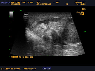
ultrasonography with color and power Doppler
imaging has emerged as the primary imaging modality for the diagnosis of
testicular torsion.It not only helps in corroborating the diagnosis by alteration of
testicular echotexture but also provides valuable information on vascular
perfusion of the testis. In addition, sonographic findings frequently allow
other diagnoses to be made in those patients presenting with an acute scrotum
who do not have torsion..
Prior to the development of high resolution, real-time
ultrasonography coupled with sensitive color Doppler, nuclear scintigraphy was
the mainstay of tests available to evaluate the acute scrotum.
Given associated
radiation, less widespread availability, limited ancillary information, and the
accuracy of color Doppler imaging, scrotal scintigraphy is no longer used as
frequently
..
Information about the role of MRI in the diagnosis of torsion
is limited.
Although MRI is likely to be highly sensitive..
However, with its limited availability, particularly at
night, and its cost, MRI is unlikely to become a front-line examination for the
patient presenting with acute scrotal pain.
Limitations of techniques
Color
Doppler ultrasonography is highly
operator dependent.
In the diagnosis of
testicular torsion, gray-scale findings are combined with dynamic flow
information. Inaccurate results may be obtained in the prepubertal patient with
small testicular volume or in cases with multiple imaging and Doppler
artifacts. Such imaging artifacts may result from inappropriate gain settings
and the non-use of slow-flow techniques..
|


No comments:
Post a Comment