Saturday, 29 September 2012

Tube within Tube-Ultrasonographic appearence of ascaris lumbricoides in small bowel

Hrsg image of fluid filled small bowel loops


Hrsg image of small bowel showing ascaris in transverse view


Ascaris lumbricoides in the gastrointestinal  tract has various features.
 Sonographic examination in the diagnosis of ascaridiasis has a high value.

The conventional method of diagnosing ascariasis is by testing the stool for the presence of the eggs.
 When there are atypical abdominal symptoms in association with a vague abdominal mass, a routine ultrasound scan using the common 3-3.5 MHz probe yields no definite findings to diagnose intestinal ascariasis.
 If a high frequency probe of -10-12 MHz is used instead, intestinal ascariasis could be definitely established.
 This case illustrates a typical worm mass diagnosed with the help of a high frequency probe.
 .

Roundworm infestation, one of the most common helminthic diseases worldwide, is caused by Ascaris lumbricoides, one of the largest parasites that infests the human bowel.
 A lumbricoides is virtually universal at some stage of childhood in semitropical and tropical regions. 

Hrsg image of small bowel showing ascaris in longitudinal view


A longitudinal section of a segment of the Ascaris lumbricoides shows the tubular shadow with brighter parallel walls (strip sign), marked by the arrows. 
The core is hypoechoic (inner tube sign). The patient's bowel walls are also identifiable.

Sunday, 16 September 2012

Polypoid Lessions in the gall bladder

polypoid lesions of the gallbladder  are defined as masses protruding from
 the mucosal surface of the gallbladder.
polypoid lessions in the gall bladder can be benign or malignant.
polypoid lessions in the gall bladder are usually diagnosed with ultrasonography as nonshadowing, fixed, solid masses
 Symptoms are unusual unless other abnormalities, such as stones, are present.
 The widespread use of US in patients with suspected gallstones and screening for other abdominal diseases has increased the detection rate of polypoid lessions in the gall bladder.


Sunday, 9 September 2012

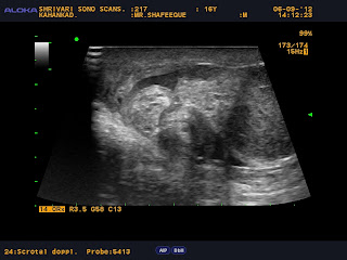
Ultrasound images in acute scrotum--Torsion Testis

Diagnostic imaging, particularly Doppler ultrasonography, plays an important role in the assessment of the patient with acute scrotal pain and swelling.
                  The affected testes was removed and found to be gangrenous
Testicular torsion, also termed torsion of the spermatic cord, is a relatively common and potentially devastating acute condition due to obstruction of the arterial blood supply to the testis.Fortunately, this entity is relatively well known, and it usually occurs with enough discomfort to lead to its diagnosis and subsequent testicular salvage
. However, atypical presentations of testicular torsion, delayed recognition of the condition, and its confusion with other causes of acute scrotum can potentially delay diagnosis and lead to testicular necrosis necessitating orchiectomy


The -"Whirl pool sign"-or-"snail sign" is also seen in the testes, caused by swollen testes  and twisted, bulky spermatic cord .
These ultrasound images suggest torsion of the Right testes.


The whirl pool sign-"snail sign" is also seen in twisted, bulky spermatic cord
(body and head of snail). 

ultrasonography with color and power Doppler imaging has emerged as the primary imaging modality for the diagnosis of testicular torsion.It not only helps in corroborating the diagnosis by alteration of testicular echotexture but also provides valuable information on vascular perfusion of the testis. In addition, sonographic findings frequently allow other diagnoses to be made in those patients presenting with an acute scrotum who do not have torsion..
Prior to the development of high resolution, real-time ultrasonography coupled with sensitive color Doppler, nuclear scintigraphy was the mainstay of tests available to evaluate the acute scrotum. 
Given associated radiation, less widespread availability, limited ancillary information, and the accuracy of color Doppler imaging, scrotal scintigraphy is no longer used as frequently
..
Information about the role of MRI in the diagnosis of torsion is limited. 
Although MRI is likely to be highly sensitive..
 However, with its limited availability, particularly at night, and its cost, MRI is unlikely to become a front-line examination for the patient presenting with acute scrotal pain.
Limitations of techniques
Color Doppler ultrasonography is highly
 operator dependent.

 In the diagnosis of testicular torsion, gray-scale findings are combined with dynamic flow information. Inaccurate results may be obtained in the prepubertal patient with small testicular volume or in cases with multiple imaging and Doppler artifacts. Such imaging artifacts may result from inappropriate gain settings and the non-use of slow-flow techniques..

Friday, 7 September 2012

Thyroid Gland Images in Acute Thyroiditis

Thyroiditis is a generic term including an array of clinical entities affecting the thyroid gland.
The thyroiditis may present infectious, autoimmune, traumatic, postradiotherapy, drugs use, and other etiopathogenies.


Heterogenous goitre with parenchymal micronodules,poorly defined hypoechoic areas permiated by fibrous echogenic layers giving a gland a psuedolobular appearece.


Doppler mapping showing a  diffuse hypervascularization pattern


Tuesday, 4 September 2012

Baker's cyst--Popliteal cyst.

Baker's cyst is a swelling that can develop behind the knee. It is filled with synovial fluid which is the lubricating fluid that is usually found inside the knee joint. It most commonly occurs if there is an underlying problem with your knee such as osteoarthritis

A Baker's cyst is a fluid-filled swelling that can develop behind the knee. It is named after a doctor called William Baker who first described this condition in 1877. It is also sometimes called a popliteal cyst, as the medical term for the area behind the knee is the popliteal fossa.


Symptoms can include pain, swelling and tightness behind the knee. Rarely, a Baker's cyst can rupture (split open) and cause similar symptoms to a deep vein thrombosis. A Baker's cyst often gets better and disappears by itself over time. However, there are various treatments that may help if you do have symptoms associated with it.

Hrsg imaging scrotum-Images of epididymis in Acute Epididymitis

The epididymis is a coiled tubular structure located along the posterior aspect of the testis. It allows for the storage, maturation, and transport of sperm, connecting the efferent ducts of the testis to the vas deferens.

Inflammation of the epididymis is much more common than orchitis. Epididymitis usually arises as a complication of an urinary system infection and is usually caused by a bacteria.


Note the free fluid collection with internal flimsy septations

On colour doppler --reveals incresed vascularity

Enlarged edematous epididymis