Sunday 9 December 2012

Ventral Hernia with obstruction-(ventral hernia specified as incarcerated,irreducible,strangulated-or-causing obstruction

ventral hernia 
Hrsg abdomen obtained with the patient at rest shows ventral hernia containing omental fat

Hrsg abdomen during valsalva maneuver reveals the hernia contains small bowel loops .
Increased intra abdominal pressure may improve the detection of small abdominal wall defect.


Incarcerated hernia

Note the bowel wall thickening, severe fat stranding, mesenteric engorgement, and extraluminal fluid confined to the hernia sac, findings that suggest incarceration.(strangulation)

Hrsg image of the abdomen shows herniation of bowel loops(multiple arrows) through a narrow abdominal wall defect. The patient was asymptomatic but presented with acute abdomen.Hrsg image shows ventral hernia (arrowheads). The sac of the hernia shown in now contains extraluminal fluid ,mildly thickened bowel loops and causes small bowel obstruction.
 Incarceration with small bowel obstruction was confirmed at surgery.

Saturday 1 December 2012

Opthalmic ultrasound- endopthalmitis(pyo-opthalmos)

Endopthalmitis is a catastrophic intraocular inflammation with or without infection occuring as a complication of intraocular surgery,trauma or uveitis.

Pain,redness and decresed vision are the most common presenting symptoms.

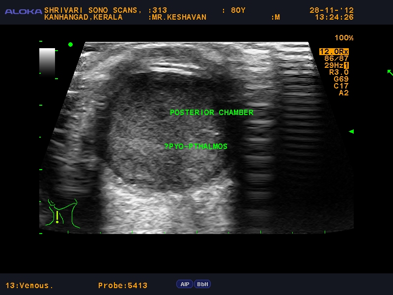
Fundal  view may be hazy or absent.

Ultrasonography is indicated to determine the presence of inflammation in the vitreous cavity and diagnose associated findings.serial ultrasounds are indicated to assess the response to both conservative treatement as well as surgical intervention..
Here the patient presented with pain ,redness and decresed vision following cataract surgery.

HRSG shows high reflective membrane like  vitreous echoes .

Hrsg right eye shows dense homogeneous low level collection filling the posterior segment with edematous eye coat suggestive of pyogenic collection.

Hrsg left eye shows Retinal detachment-it  is a disorder of the eye in which the retina peels away from its underlying layer of support tissue. Initial detachment may be localized, but without rapid treatment the entire retina may detach, 
leading to blindness.


Retinal detachment--Retinoschisis.
HRSG shows a typical echogenic peeled membrane in the posterior segment of the eye .


Hrsg shows dense homogeneous low level collection filling the posterior segment with edematous eye coat and thickened partially opaque lens displaced in posterior segment.(partially absorbed dislocated lens seen in the posterior segment)