Friday 31 August 2012

Ultrasonography for the diagnosis of appendicitis..

Diagnostic Imaging: Ultrasound: Findings in Appendicitis.

The sonographic diagnosis of acute appendicitis is based on identification of a tubular, noncompressible, aperistaltic bowel loop, which demonstrates a connection with the cecum and a distal blind end, with a diameter greater than 6 mm


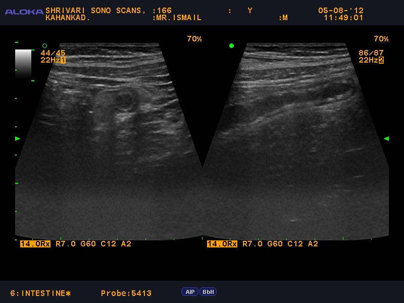
ultrasound of the right lower quadrant with the appendix showing the "target" sign seen in acute appendicitis..
Graded compression ultrasonography is one of the two imaging modalities commonly used in the assessment of 
clinically suspected appendicitis..
This self-localization technique has a sensitivity of 85%, specificity of 60%, and accuracy of 75% in diagnosing appendicitis. The overall accuracy of this technique in diagnosing appendicitis as well as other significant disease processes which may clinically mimic appendicitis is 86%. However, the self-localization technique is of limited value with retrocecal or perforated appendicitis due to the patient's inability to accurately localize the pain (21).
Based on the data of several recent studies, CT performed for clinical suspicion of appendicitis with atypical symptoms has a sensitivity of 87 - 96%, specificity of 89 - 97%, positive predictive value of 95%, negative predictive value of 95%, and accuracy of 94% (27,29).


Perforated appendix with local peritonitis

inflammed appendix with echogenic edematous omentum around

acute appendicitis

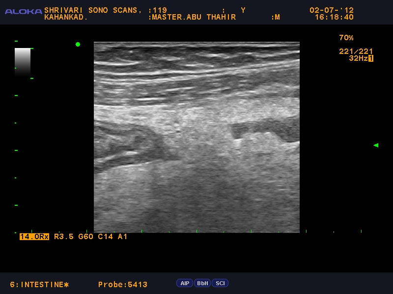
Retrocaecal appendix


retrocaecal appendix showing target sign.

Inflammed tip of the appendix.

Wednesday 29 August 2012

Enlarged Para aortic and Ileac Group of Lymphnodes-LYMPHOMA

65 years old man c/o vauge abdominal pain and bloating-h/o constipation since 2 months
Enlarged lymphnodes--
This case is biopsy proven follicular lymphoma.  Lymphoma is a neoplastic disease originating from the cells of the immune system; it typically arising within lymph nodes.
Lymphoma can involve all organ systems and site including the abdomen, thorax, pelvis, head and neck, CNS and bone.


Diagnostic imaging - including conventional radiography, US, CT and MR - plays an important role in the diagnosis and staging of lymphoma.  Sonographic evaluation is typically included in the initial workup of patients with abdominal symptoms. Therefore familiarity with the spectrum of US findings associated with lymphoma is essential to making the diagnosis.
  • Lymph Nodes-The sonographic appearance of lymphoma is variable, but hypoechoic or anechoic lymph nodes without distal enhancement are typical.  Nodal involvement can be individual or in groups.  Nodal masses may be seen adjacent to the abdominal great vessels, within the retroperitoneum, the prevertebral area and mesentery.  Lymph node encasement of the root of the mesentery and SMA may produce the “sandwich sign”, which is created by the lobulated, confluent mesenteric soft-tissue masses and the tubular structures of the mesenteric vessels and perivascular fat.

  • GI tract- Lymphomatous involvement of the bowel produces hypoechoic bowel wall thickening or a focal hypoechoic/complex mass.

  • Spleen- Sonographic findings of splenic involvement can include homogeneous splenomegaly and/or solitary nodules that are hypoechoic in comparison to the normal splenic parenchyma.

  • Liver- Hepatic US findings are similar to those of the spleen and include hepatomegaly and hypoechoic/anechoic nodules

  • Pancreas- Pancreatic involvement may appear as hypoechoic areas of enlargement.  Diffuse enlargement of the pancreas has been reported, possibly due to direct infiltration.  Peripancreatic lymph nodes may invade or distort the pancreas. 

  • Kidneys- Renal US findings can appear as hypoechoic/anechoic nodules which may be mistaken for renal cysts; however, there is a lack of distal enhancement suggesting that the mass is solid.              

In this case, further evaluation with Contrast Enhanced CT was performed. It shows multiple moderately enlarged mesenteric lymph nodes which correlate with the ultrasound findings above.

Tuesday 28 August 2012

Hrsg imaging of epigastric hernia.

Ultrasound images show the large midline defect in the abdominal wall with hernial sac protruding through the epigastric defect:


Small bowel intussusception secondary to inflammatory fibroid polyp of the ileum


Inflammatory fibroid polyps are rare benign lesions of uncertain origin,
That may occur in many different locations in the gastrointestinal tract, the gastric antrum being the most common site, followed by the small bowel.
 These lesions can cause abdominal pain, gastrointestinal bleeding, intestinal obstruction or intussusception. In the case of a gastric inflammatory fibroid polyp in the presence of Helicobacter Pylori infection, the patient can benefit from pharmacological eradication of Helicobacter Pylori.

 In case of an intestinal inflammatory fibroid polyp causing acute abdomen, the treatment is surgical.

In This case during  surgery an ileal tumour 15 cm from the ileocecal valve causing ileoileal intussusception was found.
 The intussuscepted segment was resected and an end-to-end anastomosis was carried out

Sunday 19 August 2012

scrotal hrsg --reveals testicular fracture


scrotal ultrasonography shows a fractured  testicle demonstrating discontinuity of the tunica albuginea and extrusion of testicular parenchyma .
 Mixed echogenicity from an intratesticular hemorrhage and a scrotal hematoma are also visible


biology of prenatal devalopment 8-9 weeks pregnancy

early pregnancy images-
 image gallery--prenatal devalopment -early embryo of 8-9 weeks
tiny limb buds

embryonic cardiac pulsations

the embryo is very active at times which is essential for the devalopment and maintaining healthy  muscles and bones


Wednesday 15 August 2012

fetal abdominal wall septated cystic lymphangioma diagnosed prenatally at 22 weeks

fetal chest and abdominal  wall cystic lymphangioma diagnosed prenatally at 22 weeks’ gestation is reported

.
Ultrasound examination showed a multilocular, large cystic mass (10 × 22 × 29 mm) on the left side of the fetus in the area of the lower chest and upper abdomen, without color flow imaging--ongoing preg.along with syndactyle--diagnosed 3 days back.


Saturday 11 August 2012

patent vesico urachal diverticulum


10 years old child c/o pain abdomen and  recurrent urinary tract infection --

--abdominal ultrasonography --with hfp-revealed --patent vesico urachal diverticulum--