Sunday 9 December 2012

Ventral Hernia with obstruction-(ventral hernia specified as incarcerated,irreducible,strangulated-or-causing obstruction

ventral hernia 
Hrsg abdomen obtained with the patient at rest shows ventral hernia containing omental fat

Hrsg abdomen during valsalva maneuver reveals the hernia contains small bowel loops .
Increased intra abdominal pressure may improve the detection of small abdominal wall defect.


Incarcerated hernia

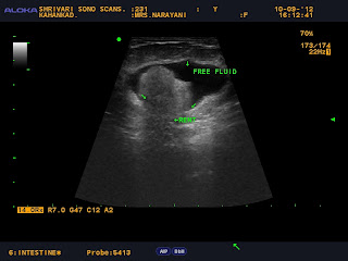
Note the bowel wall thickening, severe fat stranding, mesenteric engorgement, and extraluminal fluid confined to the hernia sac, findings that suggest incarceration.(strangulation)

Hrsg image of the abdomen shows herniation of bowel loops(multiple arrows) through a narrow abdominal wall defect. The patient was asymptomatic but presented with acute abdomen.Hrsg image shows ventral hernia (arrowheads). The sac of the hernia shown in now contains extraluminal fluid ,mildly thickened bowel loops and causes small bowel obstruction.
 Incarceration with small bowel obstruction was confirmed at surgery.

Saturday 1 December 2012

Opthalmic ultrasound- endopthalmitis(pyo-opthalmos)

Endopthalmitis is a catastrophic intraocular inflammation with or without infection occuring as a complication of intraocular surgery,trauma or uveitis.

Pain,redness and decresed vision are the most common presenting symptoms.

Fundal  view may be hazy or absent.

Ultrasonography is indicated to determine the presence of inflammation in the vitreous cavity and diagnose associated findings.serial ultrasounds are indicated to assess the response to both conservative treatement as well as surgical intervention..
Here the patient presented with pain ,redness and decresed vision following cataract surgery.

HRSG shows high reflective membrane like  vitreous echoes .

Hrsg right eye shows dense homogeneous low level collection filling the posterior segment with edematous eye coat suggestive of pyogenic collection.

Hrsg left eye shows Retinal detachment-it  is a disorder of the eye in which the retina peels away from its underlying layer of support tissue. Initial detachment may be localized, but without rapid treatment the entire retina may detach, 
leading to blindness.


Retinal detachment--Retinoschisis.
HRSG shows a typical echogenic peeled membrane in the posterior segment of the eye .


Hrsg shows dense homogeneous low level collection filling the posterior segment with edematous eye coat and thickened partially opaque lens displaced in posterior segment.(partially absorbed dislocated lens seen in the posterior segment)

Friday 23 November 2012

Opthalmic ultrasound-Retinopathy of prematurity(ROP)

Retinopathy of prematurity is seen in low birth weight premature infants due to abnormal postnatal growth of retinal vasculature.
ULTRASONOGRAPHY HELPS TO DIFFERENTIATE IT FROM OTHER CAUSES OF LEUKOCORIA.
Ultrasonography reveals bilateral small sized eyes with funnel shaped high reflective membrane attached to the optic disc suggestive of total retinal detachment..


Axial B scan image shows funnel shaped highly reflective membrane inserting into the optic disc.
Accumulation of echoes in the anterior part suggesting the presence of preretinal fibrous tissue.


Axial B scan reveals the exact configaration of the funnel

The B scan image shows packed retrolental echoes with retinal detachment

Wednesday 21 November 2012

MSK-HRUS-Forearm intramuscular venous malformation with phlebolith. .

MSK HRUS volar aspect of right forearm reveals an ill defined oval hypoechoic subcuateneous non tender sponge like mass containing intramuscular network of anechoic channels with a well defined hyperechoic mass casting posterior shadow .
left fore arm flexor muscle appears normal.


The mass lession confined to the subcutaeneous plane with a hyperechoic mass casting posterior shadow-s/o phlebolith .
Here the radiographic examination revealed rounded echogenic calcified lession.


Colour doppler application shows few week signals of flow
within the soft tissue mass.


OPTHALMIC ULTRASOUND.Retinopathy of prematurity(ROP)

OPTHALMIC ULTRASOUND.Retinopathy of prematurity(ROP)

Retinopathy of prematurity is seen in low birth weight premature infants due to abnormal postnatal growth of retinal vasculature

ULTRASONOGRAPHY HELPS TO DIFFERENTIATE IT FROM OTHER CAUSES OF LEUKOCORIA.
Ultrasonography reveals bilateral small sized eyes with funnel shaped high reflective membrane attached to the optic disc suggestive of total retinal detachment..

Axial B scan image shows funnel shaped highly reflective membrane inserting into the optic disc.accumulation of echoes in the anterior part suggesting the presence of preretinal fibrous tissue

Axial B scan reveals the exact configaration of the funnel 

The B scan image shows packed retrolental echoes with retinal detachment.

Monday 19 November 2012

Nuchal cord---Cord around the neck

Nuchal cord -or- Cord around the neck occurs when the umbelical cord becomes wraped around the fetal neck 360 degrees.
Nuchal cords are very common, the incidence of nuchal cord

 increases with advancing gestation from 12% at 24 to 26 

weeks to 37% at term
Most are not associated with perinatal morbidity and mortality.In some fetuses and newborns CAN may cause problems, especially when the cord is tightly wrapped around the neck

The cluster of cardiorespiratory and neurological signs and symptoms associated with unique physical features that occur secondary to tight cord-round-the-neck has been referred to as 'tCAN syndrome' (tight Cord Around the Neck Syndrome) 
A small number of studies have shown that nuchal cord and or tCAN can affect the outcome of delivery and may have long-term effects on the infant  and but as a causative factor for stillbirth it is debatable .


Nuchal cord twice around the neck


It is the unique physical features of tCAN syndrome that 

distinguishes it from birth asphyxia even though there are many 

similarities between these two conditions. 

Umbilical cord abnormalities are considered as one of the 

causative factor for birth asphyxia. 

The manifestation of tCAN symptomatology seems to happen both 

in the presence of normal and depressed AGPAR scores. 

Umbilical cord compression due to tCAN may cause 

obstruction of blood flow first in thin walled umbilical vein, while

 infant’s blood continues to be pumped out of baby through the

 thicker walled umbilical arteries thus causing hypovolemia and

 hypotension resulting in acidosis . Anemia  and mild

 respiratory distress may occur. Some of these infants may also 

have facial and conjuctival petechiae and rarely petechiae of

 the neck and upper part of the chest and skin abrasion of neck

 where the cord was tightly wrapped and facial suffusion 

 all of which can also be seen in some postmortem findings of 

stillbirth infants who had tCAN.

 If born alive, some of these infants may also be

 somewhat obtunded with a low tone and have transient feeding 

difficulties. These findings raise the possibility of transient

 encephalopathy, which may lead to long-term complications.

Friday 9 November 2012

Intrauterine synechiae-Amniotic sheets visualized in the uterus

The differential diagnosis of reflective intrauterine membranes during pregnancy includes amniotic bands of fetal origin or membranes of combined maternal-fetal origin. While the former have been associated with fetal transverse reduction defects or the lethal amniotic band syndrome, the latter are usually benign and consist of a preexisting uterine septation or synechia around which the fetal membranes fold or become enveloped.

Intra Uterine Synechiae:
(also known as "amniotic sheets" or "amniotic folds")

A synechia is a term meaning an "adhesion", or a fibrous scar. Uterine synechiae have also been referred to as "amniotic sheets" or "amniotic folds". 

Many patients with sonographically demonstrable uterine synechia have clinical history 
of uterine instrumentation or uterine infections to explain scar formation. Most commonly, uterine synechiae are noted as an incidental finding on obstetric sonograms.

 In general, synechiae do not interfere with the development and growth of pregnancy, 
and are rarely associated with any complications.  There is some evidence to suggest that large synechiae may be the cause of malpresentation due to partial compartmentalization of the uterine cavity, and may also be associated with lower birth weight.
Visualization of a band-like structure should not be confused with amniotic band syndrome especially when thorough fetal anatomy survey reveals no structural 
anomalies.

fetal face .nose and lip seen.

normal fetal hand and wrist seen.

coronal view of fetal face with upper limb


Here We present an unusual case in which a patient was noted at 30 weeks' gestation to have an 

intrauterine membrane of undetermined origin, appearing to contain a pulsating vessel


Color Doppler imaging and Doppler flow velocimetry clearly depicted a pulse synchronous with the

maternal heart rate, defining the membrane as amniotic membranes surrounding a preexisting

uterine synechia and not a true amniotic band. 


There is some evidence to suggest that large synechiae may be the cause of 

malpresentation due to partial compartmentalization of the uterine cavity.


There were no obvious structural anomalies seen.


Visualization of a band-like structure should

 not be confused with amniotic band syndrome especially when thorough fetal anatomy

 survey reveals no structural anomalies.


There is some evidence to suggest that large synechiae may be the cause of 

malpresentation due to partial compartmentalization of the uterine cavity 

Here the fetus is of 30 weeks gestation  in transeverse lie

Visualization of a band-like structure should not be confused with amniotic band 

syndrome especially when thorough fetal anatomy survey reveals no structural 

anomalies.